Medial Epicondylitis (Golfer's Elbow)
Medial epicondylitis, or “golfer's elbow,” typically arises from repetitive motion — most commonly gripping and grasping. With continuous, forceful flexion of the wrist, the tendons of the forearm flexor muscles become overworked and develop small tears and inflammation. Symptoms are mostly on the inside of the elbow and along the palm side of the forearm. Diagnosis is usually made by physical exam, although an MRI can be helpful.
Treatment is usually non-surgical: reducing inflammation with oral anti-inflammatories or a cortisone injection, and stretching the affected muscles. Physical therapy helps, and braces are available for compression and support.
While this can be a stubborn condition, most patients do well with conservative care. If symptoms continue despite appropriate treatment, surgical debridement is an option. PRP (platelet-rich plasma) injections are another treatment option for chronic cases.
Lateral Epicondylitis (Tennis Elbow)
Lateral epicondylitis, or “tennis elbow,” typically arises from repetitive grasping, pushing, pulling and lifting. With continuous, forceful extension of the wrist, the tendons of the forearm extensor muscles become overworked and develop small tears and inflammation. Symptoms are mostly on the outside of the elbow and can extend into the forearm. Diagnosis is usually made by physical exam, although an MRI can be helpful.
Treatment is usually non-surgical: reducing inflammation with oral anti-inflammatories or a cortisone injection, and stretching the extensor muscles. Physical therapy helps, and braces are available for compression and support.
Most patients do well with conservative care. If symptoms continue, surgical debridement is an option. PRP (platelet-rich plasma) injections are another treatment option for chronic lateral epicondylitis.
Cubital Tunnel Syndrome
Cubital tunnel syndrome is compression of the ulnar nerve across the elbow. It can result from direct trauma, constant or repetitive pressure over the nerve, or prolonged or repeated bending of the elbow.
Although the problem is at the elbow, most symptoms are felt in the hand — typically numbness and tingling in the ring and small fingers. Patients may notice decreased grip strength, reduced coordination when gripping and grasping, and decreased sensation. With severe or long-standing compression, muscle wasting in the hand can occur.
Diagnosis is made clinically, with MRI and nerve conduction studies as helpful additions. Mild symptoms can be managed with anti-inflammatories, physical therapy and bracing. If muscle wasting has occurred, prompt surgical release is recommended to prevent further loss of nerve function — opening the cubital tunnel, freeing the nerve from scar tissue, and repositioning it to avoid recurrent compression.
Olecranon Bursitis
The olecranon bursa is a small fluid-filled sac under the skin at the tip of the elbow that provides cushioning and lubrication. Inflammation of the bursa — bursitis — can result from trauma, prolonged pressure, or infection. It typically appears as painful swelling over the elbow; range of motion is generally unaffected. Redness and tenderness may indicate infection.
Diagnosis is made by physical exam; MRI is typically not needed. X-rays are taken to rule out a bone spur on the olecranon, which can predispose you to bursitis. Treatment involves compression, ice, activity modification and anti-inflammatory medication.
Most often, the bursa is aspirated to remove accumulated fluid, and a small amount of cortisone is injected to reduce inflammation and help prevent recurrence. If infection is present, antibiotics are started after aspiration. Chronic cases may need repeat aspiration, and surgical removal of the bursa is available, though most patients improve with aspiration alone.
Radial Head & Neck Fractures
Radial head and neck fractures account for about 20% of elbow injuries, most often after a fall. Symptoms include pain over the outside of the elbow, swelling, bruising, difficulty moving the elbow, and pain when rotating the forearm. X-ray and CT confirm the diagnosis.
These fractures are classified by size, location and displacement. Most can be treated conservatively with short-term immobilization, ice and rest, followed by physical therapy. Depending on the nature of the fracture, surgery may be indicated.
Elbow Dislocations
Elbow dislocations are uncommon and often result from a fall onto an outstretched hand. They are categorized as simple (no bone injury), complex (bone and ligament injury) or severe (injury to nearby nerves and vessels). X-ray and CT confirm the diagnosis.
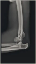
The injury causes pain and obvious deformity. The joint must be manually reduced, usually in an emergency room or urgent care setting.
Simple dislocations are treated with short-term sling immobilization followed by early range-of-motion exercises with a physical therapist. Some patients may not regain full extension, though the elbow still functions well with this limitation. Complex and severe dislocations most often require surgery to stabilize and repair bone and ligament injuries.