Rotator Cuff Tears
Tears of the rotator cuff can happen in a variety of ways. They can result from repetitive trauma or a single injury, or they can be degenerative. Depending on your symptoms and the severity of the tear, treatment can vary.
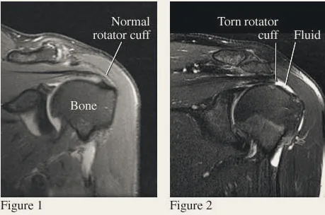
The rotator cuff is made up of the tendons of four muscles inside the shoulder. These muscles are responsible for the stability and function of the shoulder joint. Symptoms associated with tearing include pain in the front and outside of the shoulder, pain radiating to the upper arm, weakness, and nighttime pain. Diagnosis is confirmed with a good-quality MRI.
Treatment involves both surgical and non-surgical options. Symptomatic relief can be found with oral anti-inflammatories or a cortisone injection, an ice regimen and physical therapy. If the tear is small, these measures are often all you need to return to activity. Larger or recurrently symptomatic tears may require surgery.
Surgery is done arthroscopically through three or four small incisions. The rotator cuff is reattached to its footprint on the humerus with small suture anchors that allow the tendon to heal over time. An acromioplasty, or decompression, is also performed to give the rotator cuff more room to function. Recovery involves sling immobilization for approximately four to six weeks, followed by physical therapy to regain motion and strength.
Shoulder Impingement
Like rotator cuff tears, shoulder impingement can occur in a variety of ways — most often from repetitive trauma. Under the acromion bone is a small bursal sac that overlies the rotator cuff. A down-sloping acromion, or a bone spur, reduces the space for the bursa and rotator cuff. Combined with repetitive activity, this places you at risk of impingement.
Symptoms include pain in the front and outside of the shoulder when activities are performed at or above shoulder level. An MRI is helpful in establishing the diagnosis and ruling out rotator cuff tearing.
Treatment focuses on reducing bursal inflammation with oral anti-inflammatories or a cortisone injection. Physical therapy is also recommended to reduce inflammation and strengthen the shoulder.
Occasionally, impingement becomes chronic and recurring. In that case, an arthroscopic decompression of the acromion with debridement of the bursa can be performed.
Shoulder Labral Tears
The shoulder is a ball-and-socket joint held together in part by the labrum — a ring of tissue and ligaments that provides stability. Labral tears can occur through traumatic dislocation, repetitive trauma, or a single injury.
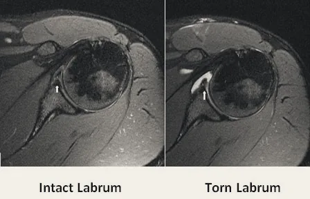
Symptoms can include frank instability or a sensation of looseness in the joint, vague achiness or discomfort, and clicking or catching inside the shoulder. Diagnosis is confirmed with an MRI arthrogram.
Treatment involves both surgical and non-surgical options. Conservative treatment focuses on reducing inflammation and strengthening the joint with physical therapy. Depending on symptoms and the nature of the tear, surgical repair with suture anchors may be necessary to fix the labrum back to the bone.
Shoulder Dislocation & Instability
The shoulder has the greatest mobility of any joint — and with that mobility comes a risk of instability. Instability can result from a traumatic dislocation or progress slowly with repeated stress. Tears of the labrum and surrounding ligaments can cause chronic, recurrent instability.
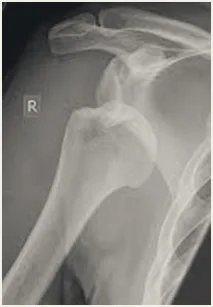
Traumatic instability following a dislocation involves a tear of the labrum from the socket. In young and athletic patients especially, recurrent dislocation is common. Treatment initially aims to reduce inflammation and restore motion. Should conservative measures fail and instability recur, surgical reconstruction of the labrum is a good option.
Minimally invasive arthroscopic surgery reattaches the torn labrum to the glenoid using small suture anchors, allowing it to heal and re-stabilize the shoulder. Recovery involves sling immobilization for about four weeks, followed by dedicated physical therapy to regain motion and strength.
Patients with non-traumatic instability — from repetitive stress or naturally loose ligaments — rarely have labral tears. Treatment in this case focuses on stabilizing and strengthening the shoulder with physical therapy.
Shoulder Arthritis
Shoulder osteoarthritis is wear and tear of the cartilage covering the ball and socket. As the cartilage continues to wear, the joint eventually develops bone-on-bone contact. The most common symptoms are pain and stiffness.
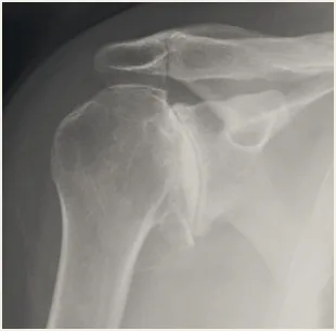
Once cartilage is lost, it cannot be replaced. Symptoms can be managed with medication and physical therapy to reduce pain and improve function. Ultimately, a shoulder replacement may be needed.
There are two approaches: a traditional shoulder replacement and a reverse shoulder replacement. The surgical technique is similar; the right option depends on the integrity of your rotator cuff. Patients with an intact rotator cuff can proceed with a traditional replacement. Patients with a chronically torn rotator cuff require a reverse replacement, in which the positions of the ball and socket are reversed, removing the need for a functioning rotator cuff.
With either procedure, recovery involves six weeks of sling immobilization followed by physical therapy to regain range of motion and strength.
Frozen Shoulder
Frozen shoulder, or adhesive capsulitis, is a painful stiffening of the shoulder capsule that restricts range of motion. It often occurs without a specific cause, although it can be associated with trauma. It is most common between the ages of 40 and 60 and has some association with diabetes and thyroid conditions.
Symptoms include progressive stiffness and pain. Diagnosis is made by physical exam; an MRI can help rule out other issues but is not usually necessary.
Treatment involves dedicated physical therapy to stretch the capsule and regain motion, and a cortisone injection to reduce inflammation, pain and scar tissue. Often, therapy and an injection alone resolve the problem.
For refractory cases, two procedures are available. A manipulation under anesthesia gently moves the shoulder through its range of motion while you are asleep, breaking up the tight, scarred capsule. If this fails, an arthroscopic release uses three small incisions and a camera to open the capsule directly, allowing it to heal in a lengthened position. Physical therapy follows either procedure to keep the capsule from tightening again.