Knee Arthritis
Arthritis is most common in weight-bearing joints such as the knee. It is wear and tear of the joint cartilage, narrowing the joint space and causing pain and stiffness. It develops gradually, and symptoms often progress with it. Diagnosis is confirmed with X-ray.
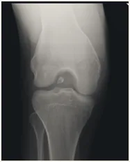
Patients may also experience nighttime discomfort, loss of motion, and difficulty walking long distances. There is no cure for arthritis; once cartilage is damaged, it cannot be replaced.
Conservative options include anti-inflammatory medication, periodic cortisone injections into the joint, and physical therapy to regain motion and strength.
Surgical treatment is a knee replacement, which removes the arthritic joint surfaces and replaces them with an artificial joint. Physical therapy follows to regain motion and strength.
Meniscus Tears
Each knee has two menisci — medial and lateral — washer-like structures that sit between the bones of the knee and provide cushioning. Meniscus tears are very common and often follow a twisting injury.
Symptoms include swelling, popping, catching, clicking, pain and sometimes restricted motion. Fully bending the knee is often painful, as is twisting or pivoting. Diagnosis is confirmed with an MRI.
The meniscus has a limited blood supply, so most tears do not heal on their own. Initial treatment reduces inflammation, pain and swelling and restores motion. Most meniscus tears, however, do require surgery — performed arthroscopically to remove the torn portion. Occasionally a meniscus repair is possible, reserved for tears near the meniscus's attachment to the bone. The type of tear and surgical recommendation depend on MRI findings.
ACL Tears
The anterior cruciate ligament (ACL) is a major stabilizing ligament in the center of the knee. ACL tears typically result from a high-energy movement — changing direction rapidly, stopping suddenly, landing awkwardly from a jump — or from direct contact. A loud “pop” is often heard.
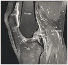
Pain and swelling usually appear within 24 hours, accompanied by instability. Once the acute swelling resolves, many patients feel well, but the knee remains unstable and a return to previous activities is not possible. Diagnosis is made by physical exam and MRI.
ACL tears do not heal on their own. Depending on age and activity level, surgery is usually needed. It is performed arthroscopically, reconstructing the ligament with either a donor graft or the patient's own hamstring tendon. Other injuries, such as meniscus tears, are addressed at the same time.
Patellofemoral Pain Syndrome
Patellofemoral pain is a general term for pain around the kneecap (patella), often called “jumper's knee” or “runner's knee.” It is common in athletes and slightly more common in young adults and women. The most common symptom is pain at the front of the knee that makes climbing stairs, bending, squatting and kneeling difficult.
It can develop from repetitive stress, a sudden change in activity frequency, duration or intensity, or improper training technique — often poor stretching. It can also result from malalignment, where the patella does not sit centered in its groove and does not track smoothly.
The most common contributor to poor tracking is muscle imbalance, tightness or weakness — particularly of the quadriceps, which help keep the patella in its groove as the knee bends and straightens.
This can be a frustrating problem that takes time to improve. Treatment focuses on reducing inflammation and stretching the quadriceps, with physical therapy and a dedicated home exercise program. Ongoing maintenance usually helps keep symptoms from recurring. Surgical options exist for severe malalignment, though they are not commonly needed.
MCL Tears & Sprains
The medial collateral ligament (MCL) is one of two collateral ligaments that provide side-to-side stability to the knee, and it is injured far more often than the lateral collateral ligament (LCL).
The MCL is usually injured by force against the outside of the knee, causing a sprain or tear. Symptoms include pain and swelling on the inside of the knee, and often instability. Diagnosis is confirmed by physical exam and often an MRI.
Treatment is conservative, as this ligament heals well on its own. A supportive knee brace is recommended for the first four weeks, along with physical therapy to regain strength and range of motion and reduce pain.
Patellar Fractures & Dislocations
Two common traumatic injuries to the kneecap are fracture and dislocation. Patellar fractures typically follow a direct fall onto the knee. Stable, non-displaced fractures can generally be treated with brace immobilization followed by gentle range of motion. Unstable or markedly displaced fractures require surgery to reduce and stabilize the fracture with hardware. Symptoms include swelling, bruising, and inability to bend or straighten the knee; diagnosis is confirmed by X-ray.
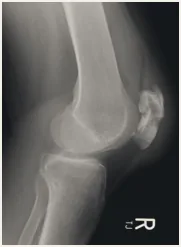
Patellar dislocations usually follow high-energy trauma. The patella slides out of its groove toward the outside of the knee, causing visible deformity; it is reduced manually, usually in an emergency room or urgent care setting. Treatment then involves brace immobilization followed by physical therapy.
A dislocation injures the medial patellofemoral ligament (MPFL) on the inside of the knee. It heals on its own but often in a lengthened position, which increases the risk of recurrent dislocation. If dislocations recur, surgical tightening or reconstruction of the MPFL can re-stabilize the kneecap.
Cartilage Damage & Loose Fragments
Cartilage covers the surfaces of the knee joint. It can be damaged in several ways, most commonly by arthritis, which thins cartilage and can leave large defects. Localized damage in an otherwise healthy knee is usually the result of a previous injury.
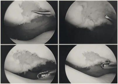
Diffuse damage causes generalized stiffness, soreness and pain. Large cartilage flaps or loose fragments can also cause mechanical symptoms — popping and catching — as they catch during movement. X-ray and MRI confirm the diagnosis.
For mechanically symptomatic cartilage damage or loose fragments, arthroscopy can smooth cartilage defects and remove detached fragments. Cartilage cannot be replaced and arthritis symptoms may persist, but popping, catching and clicking from loose fragments and flaps typically improve.